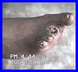
July 10th, 1996, I visited Mrs. Clara Patt. Mrs. Patt, was suffering from a severely infected right foot. Mrs. Patt had been diabetic for the past 30 years. The sixteenth of September, 1995, she developed an infected toe. On February 30, 1996, she had two toes amputated.
When I saw her July 10th, the foot was swollen, feverish, and very painful. The wound resulting from the amputation was not healing, and was a deep severely, infected, cavity. Mrs. Patt was suffering pain from the top of foot, ankle and shin. The bones of her foot were infected. Large courses of modern antibiotic treatments had been given to no effect. She was close to fatal systemic infection and the hospital wanted to proceed with amputation of entire leg below the knee immediately.
In the following "Pic" we see some of the deep cavity left from amputation and the general inflammation of her foot. As will be shown in later "Pics", this infection comes out through the bottom of the sole as well.
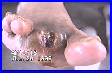
The next Picture is taken July 20, ten days later. We see that swelling is dramatically reduced. The wound is expressing it's self, the property of Cascabel treatment to self-debride is progressing as usual. Mrs. Patt no longer experiences pain and there is no longer fever present. We can also see that new flesh is being regenerated to close off this wound. Notice the infection channel breakout shown slightly on th bottom of foot.
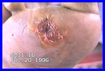
Below, we see for the first time, the wound on the bottom of the foot. This wound is one with the amputated toe's socket. This wound has debrided it's self well and is starting to regenerate flesh as well.
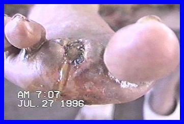
This Picture taken just seven days later of the socket wound demonstrates the rapid closing of this wound with healthy new flesh. We also see evidence of further reduction in inflammation of the entire foot.
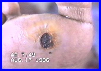
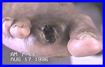
The next two Pics show the progress of treatment to August 17, 1996 -- or 20 days later. Both areas continue to heal well.
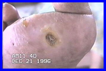
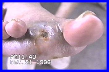
Clara Patt took her last treatment Nov. 16, 1996. The following two Pics of this same foot are taken Dec. 21, 1996 -- 35 days after her last dose of medication. The foot is well healed and stabilized.
Presented
by the work of:
Peter
Singfield
"Medicine
Man"
Xaibe Village
Corozal District,
Belize, Central America
Tel 501-4-35213